
Neonatal respiratory distress syndrome (RDS) is acute respiratory failure caused by an immature lung, principally by insufficient surfactant and incomplete gas-exchange architecture. It is the dominant early complication of and becomes more common and more severe as gestational age falls.
The abbreviation causes confusion and is worth settling at the start. Neonatal RDS is surfactant-deficient hyaline membrane disease. Adult-type acute respiratory distress syndrome (ARDS) is a different disease with a different mechanism and definition, and the two are not interchangeable even though both produce hypoxemic respiratory failure.
Why the preterm lung collapses
Lung development runs through overlapping stages. At 23 to 28 weeks the lung is in the late canalicular to early saccular phase: airways are present but the gas-exchange region is still being built. Saccular development continues to roughly 36 weeks, and alveolarization continues after birth. Surfactant is produced by type II pneumocytes and small amounts are detectable from about 22 to 24 weeks, rising substantially toward term.
Surfactant lowers surface tension at the air–liquid interface. Without enough of it, a much higher pressure is needed to open each small airspace, and unstable units collapse again at the end of expiration. The result is widespread atelectasis, reduced lung compliance, ventilation–perfusion mismatch, hypoxemia, carbon dioxide retention and respiratory acidosis. The infant spends a large amount of energy reopening the lung with every breath and tires within hours.
Transition adds a second burden. Fetal lung fluid has to be absorbed while gas exchange starts, and clearance is less efficient in the immature lung, so the first breaths have to establish aeration in a partially fluid-filled lung.
What respiratory distress looks like
An infant who has to reopen collapsing airspaces with every breath shows it as visible effort, and the signs escalate as that effort becomes harder to sustain:
- Respiratory rate above 60 breaths per minute.
- Retractions of the soft chest wall between the ribs, below the costal margin or over the sternum.
- Nasal flaring, which reduces inspiratory resistance.
- Grunting during expiration, produced by partial closure of the vocal cords. Grunting generates intrinsic positive end-expiratory pressure, a small pressure that stays in the airways at the end of expiration, and slows alveolar collapse, so it is a sign of disease that the infant is actively compensating for.
- Cyanosis, pallor, lethargy, reluctance to feed and apnea are later signs of an infant who is tiring or already failing.
Severity can be graded by scoring systems such as the , which sums the visible work of breathing. Any such score records effort, not gas exchange, so it is read together with saturation and blood gases.
Investigations
Examination shows how hard the infant is working; investigations show whether gas exchange has failed and what the lung looks like. Preductal pulse oximetry on the right hand, which measures blood that has not yet passed the ductus arteriosus (the fetal vessel between the pulmonary artery and the aorta), is combined with blood gas analysis, which may reveal hypercapnia (a raised carbon dioxide level) with respiratory acidosis.
On chest radiography, surfactant deficiency develops a reticulogranular or ground-glass pattern with , in which air-filled bronchi stand out against the airless lung around them; in the most severe the lungs become diffusely opaque and the cardiac border is hard to distinguish. Lung ultrasound is increasingly used as a bedside alternative: normal lung shows horizontal , whereas RDS produces dense vertical and, in severe disease, a “”. It can be repeated without moving a critically ill infant.
Support the lung without injuring it
The treatment logic is a ladder. An infant who breathes but cannot keep the airspaces open receives nasal continuous positive airway pressure (CPAP), which delivers distending pressure through prongs or a mask and avoids an endotracheal tube. If CPAP fails, invasive mechanical ventilation becomes necessary, but modern practice aims for the gentlest support that maintains gas exchange, because high pressures and large tidal volumes (the volume of each breath) injure the immature lung.
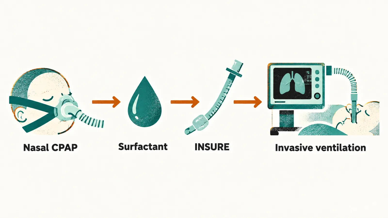
Pressure holds the airspaces open, and surfactant treats the deficiency behind their collapse, but it has to reach the lower airway. Nebulisation was not a reliable delivery route because much of the material deposits in the upper airway, so surfactant is instilled through an endotracheal tube or a thin catheter. INSURE — intubation, surfactant, extubation — describes giving surfactant through brief intubation and then removing the tube once the infant can continue on non-invasive support. An infant who already requires invasive ventilation receives surfactant through the existing tube.
Air leak is the complication that pressure can create. In a surfactant-deficient lung, some regions over-distend while others stay collapsed, and an over-distended airspace can rupture into the pleural space. Sudden deterioration in oxygenation, heart rate or blood pressure during respiratory support raises the possibility of pneumothorax, which may require immediate drainage with a chest tube. This risk is one reason to prefer CPAP and shorter periods of invasive ventilation when the clinical situation allows.
Apnea of prematurity
Not every breathing problem in a preterm infant comes from the lung. Apnea of prematurity — pauses in breathing — has a separate mechanism: the immature respiratory control center, so breathing pauses even without worsening parenchymal disease. It is common at the earliest gestations and usually improves as the infant approaches term, with respiratory control maturing around 34 to 35 weeks.
Apnea is treated with respiratory support, positioning and , which stimulates respiratory drive and reduces apneic episodes. Standard reference dosing is a loading dose of 20 mg/kg followed by maintenance of 5 mg/kg once daily, which may be increased to 10 mg/kg per day if apnea persists; the large trial that established its neonatal use also reported less bronchopulmonary dysplasia and a lower rate of death or neurodevelopmental disability at 18 to 21 months. Doses are prescribed by unit protocol rather than this note.
From acute RDS to bronchopulmonary dysplasia
Bronchopulmonary dysplasia (BPD), also called chronic lung disease of prematurity, is the chronic respiratory consequence in infants who survive immature-lung disease and prolonged oxygen or ventilatory support. Acute RDS can resolve while the infant remains dependent on respiratory support and saturation monitoring for weeks or months.
BPD is best understood as cumulative injury to a lung that is trying to grow: oxygen exposure, ventilator-induced stress, infection, inflammation, fluid overload and a persistent ductus arteriosus all contribute. Definitions vary, which is why reported incidence differs widely; among infants who survive to 36 weeks (gestational age at birth plus the weeks since), about half meet an oxygen-use definition of BPD. Severe disease prolongs the admission, delays feeding autonomy and may require discharge with home oxygen, technology dependence or, in the most severe cases, tracheostomy and long-term ventilatory support.
Both respiratory distress syndrome and BPD interact with the cardiovascular transition: lung disease promotes ductal patency, and a hemodynamically significant patent ductus arteriosus increases pulmonary blood flow and worsens the pulmonary edema that the immature lung is already struggling with. Respiratory support and feeding are connected in the same way, because an infant who is working hard to breathe has little energy budget left to feed, and the circulation that determines lung edema also determines bowel perfusion — which is the point at which the treatment of the ductus and the risk of necrotizing enterocolitis meet.