
Intraventricular hemorrhage (IVH) is bleeding that begins in the germinal matrix of the brain and reaches the ventricular system. It is the characteristic hemorrhagic injury of extreme prematurity, and its close companion, cystic periventricular leukomalacia (cPVL), is the characteristic ischemic injury of the same brain’s white matter. These two lesions are the findings that most consistently predict later cerebral palsy and cognitive difficulty in infants born at the earliest gestations.
Why this brain bleeds, and when
The germinal matrix is a thick, densely vascular region beneath the ependyma (the lining of the ventricles) beside the lateral ventricles, where neurons and glia are generated. It is at its largest between roughly 24 and 32 weeks of gestation and involutes before term. Its vessels are fragile, thinly walled and poorly supported, so they rupture more easily than mature vessels. Autoregulation, the ability of the brain to keep its blood flow steady when blood pressure changes, is also immature, which means that a sudden rise in arterial pressure, a fluid bolus, a change in carbon dioxide tension, a pneumothorax or a painful procedure can be transmitted to those vessels instead of being buffered.
The lesion is therefore an early one: most hemorrhage occurs within the first 72 hours after birth, and about half is already visible on the first day. Risk falls steeply as gestational age rises. Roughly 20% to 25% of very-low-birth-weight infants develop IVH, and the proportion with a severe bleed is highest at the earliest gestations — in one population cohort, severe hemorrhage affected about 36% of infants born at 22 to 23 weeks, 21% at 24 to 25 weeks and just under 10% at 26 to 27 weeks.
How the hemorrhage is graded
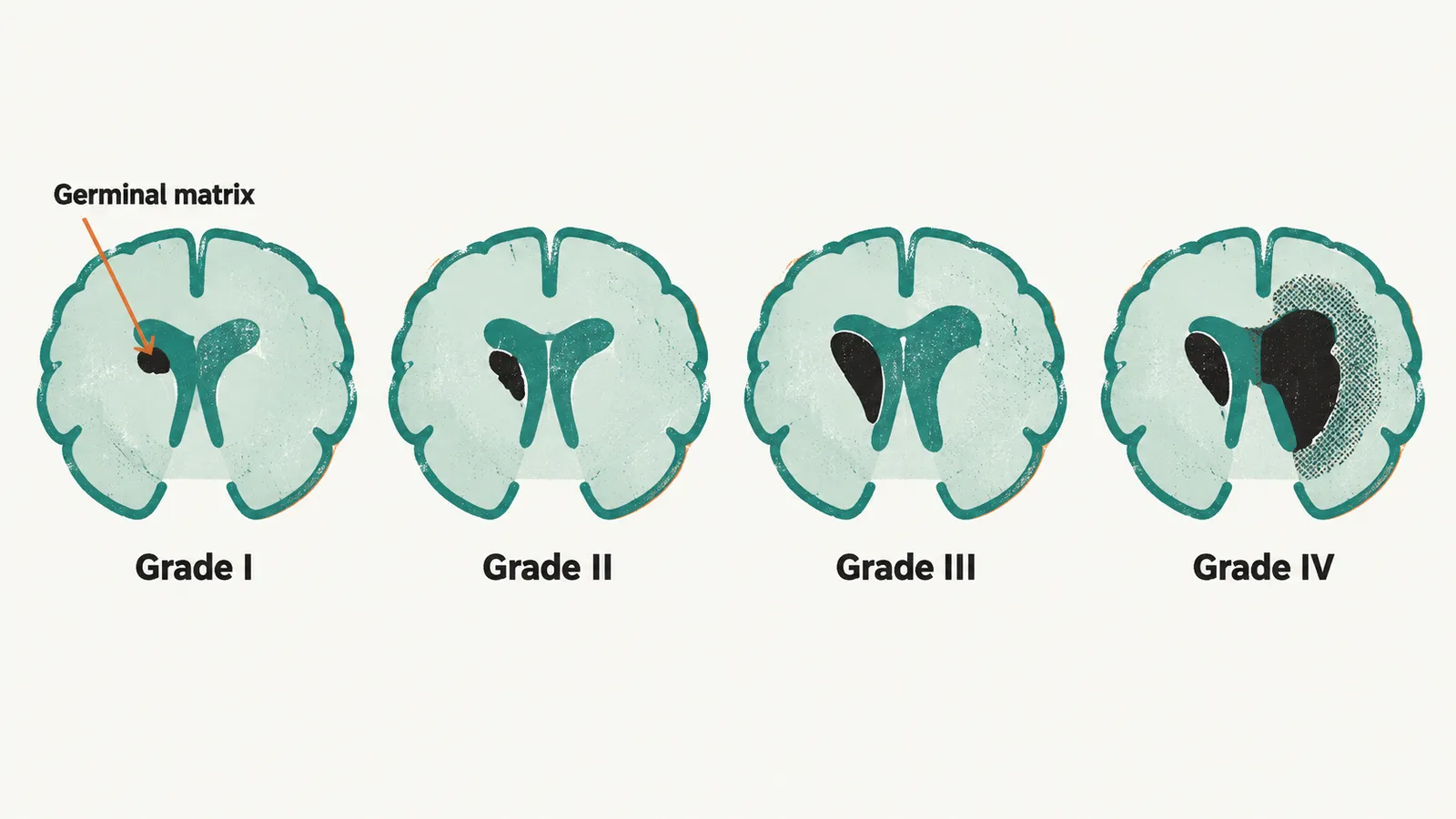
Cranial ultrasound grades the lesion, using the classification introduced by and colleagues.
| Grade | Finding |
|---|---|
| I | Blood confined to the germinal matrix (subependymal hemorrhage) |
| II | Blood within the ventricle without ventricular dilatation |
| III | Blood within the ventricle with ventricular dilatation |
| IV | Periventricular hemorrhagic infarction |
The label “grade IV” is historical in a way that matters. The parenchymal lesion it describes is now understood as a , usually a venous infarction beside the ventricle rather than blood simply spilling out of the ventricle, and it is that component — not the intraventricular blood itself — that carries the worst prognosis.
Screening before there are symptoms
An infant with IVH usually looks no different at the bedside. Apnea, a fall in hemoglobin, a bulging fontanel or a seizure appear only with large bleeds, so units scan by protocol instead of waiting for a sign. National guidance agrees on the principle and differs on the timetable.
| American Academy of Pediatrics | Canadian Paediatric Society | |
|---|---|---|
| Who is scanned | Infants born at or before 30 weeks | All infants born at or before 31 weeks and 6 days |
| First scan | By 7 to 10 days | At 4 to 7 days |
| Repeat scan | At 4 to 6 weeks | At 4 to 6 weeks |
| Later scan | At term-equivalent age or before discharge | At term-equivalent age for the most immature infants |
Two points outlive the exact dates. The first scan is done early, because the bleed happens early. And imaging is repeated, because ventricular dilatation and white-matter injury evolve after the first few days, so a normal scan in the first week does not close the question.
When blood obstructs cerebrospinal fluid
Blood in the ventricular system obstructs the flow and reabsorption of cerebrospinal fluid, and a grade III or grade IV hemorrhage may be followed by post-hemorrhagic ventricular dilatation. It is tracked by serial ultrasound measurements of the ventricles — the and the anterior horn width — rather than by head circumference or the fontanel alone, because the physical signs of raised pressure appear late in a very preterm infant.
If the ventricles continue to enlarge, cerebrospinal fluid is drained, first with a temporary device such as a ventricular access reservoir or a ventriculosubgaleal shunt, and later, in a minority of infants, with a permanent ventriculoperitoneal shunt. Serial lumbar punctures, acetazolamide or furosemide, and intraventricular fibrinolytic therapy are not recommended for preventing death or shunting, which leaves early ultrasound-guided drainage and surgery as the measures in use.
The prevention that exists
Because most bleeding happens in the first days, protection has to be in place from birth. Much of it is arranged before birth, through the obstetric management of threatened preterm delivery and through delivery in a center with neonatal intensive care, and that antenatal part of prematurity belongs with the obstetric side of the subject rather than here. In the neonatal unit, the measures available are those of gentle intensive care, and they follow from the fragile vessels and immature autoregulation described above: avoiding rapid volume expansion and swings in blood pressure, avoiding hypocarbia (a low carbon dioxide level) and large fluctuations in carbon dioxide, using the least injurious form of ventilation, keeping the head midline, and handling the infant as little as possible. Delaying cord clamping at birth, as described in The Golden Hour and NICU Care of the Preterm Infant, is associated with less intraventricular hemorrhage. The evidence for the individual postnatal measures is weaker than for the antenatal ones, and they are used together as a bundle of practices rather than as single proven interventions. The environment participates as well: light, noise, handling, pain and cardiorespiratory instability during a period that would normally be spent inside the uterus are part of the load on a brain that is still building itself, and developmental care is designed to reduce that load.
White-matter injury
Hemorrhage is not the only injury of this brain. The white matter beside the lateral ventricles is where premyelinating oligodendrocytes — the cells that will wrap axons in myelin — are concentrated during the same 24- to 32-week window. They are unusually vulnerable to low perfusion, inflammation and free-radical injury, so a fall in cerebral blood flow, a systemic infection, or the inflammatory response that accompanies (inflammatory necrosis of the immature bowel) can damage the white matter even when there is no hemorrhage at all.
The classic result is cystic periventricular leukomalacia: small cysts in the periventricular white matter that become visible on ultrasound days to weeks after the insult. Diffuse, non-cystic white-matter injury is more common than the cystic form and also affects outcome, but it is harder to see on ultrasound and may be recognized only on MRI at . Because the corticospinal tracts pass through the periventricular region, the resulting motor deficit is predominantly spastic, and cystic periventricular leukomalacia is the lesion most consistently associated with later cerebral palsy, with bilateral and extensive disease carrying the highest risk.
What the lesion predicts
Because hemorrhage and white-matter injury affect the same immature brain, prognosis has to weigh both. The grade and the location matter, and neither is a prediction of an individual child’s future on its own.
- Grade I and II hemorrhage without parenchymal involvement is associated with a measurable increase in neurodevelopmental impairment, cerebral palsy and sensory deficits compared with no hemorrhage at all — a larger increase than older teaching suggested, but much smaller than with severe bleeding.
- Grade III and IV hemorrhage, and especially periventricular hemorrhagic infarction, carries a substantially higher risk of motor impairment and of the hydrocephalus that may follow.
- Cystic periventricular leukomalacia carries the highest motor risk and is often accompanied by cognitive, visual and language difficulty.
Cerebral palsy is more common the more immature the birth, and both IVH and white-matter injury contribute to that excess risk. Outcome is never decided by the ultrasound label alone: the extent and laterality of the lesion, later imaging, seizures, hearing and vision, the family and social environment, and the child’s own developmental trajectory all contribute. That is why high-risk developmental follow-up and early therapy matter as much as the neonatal scan.