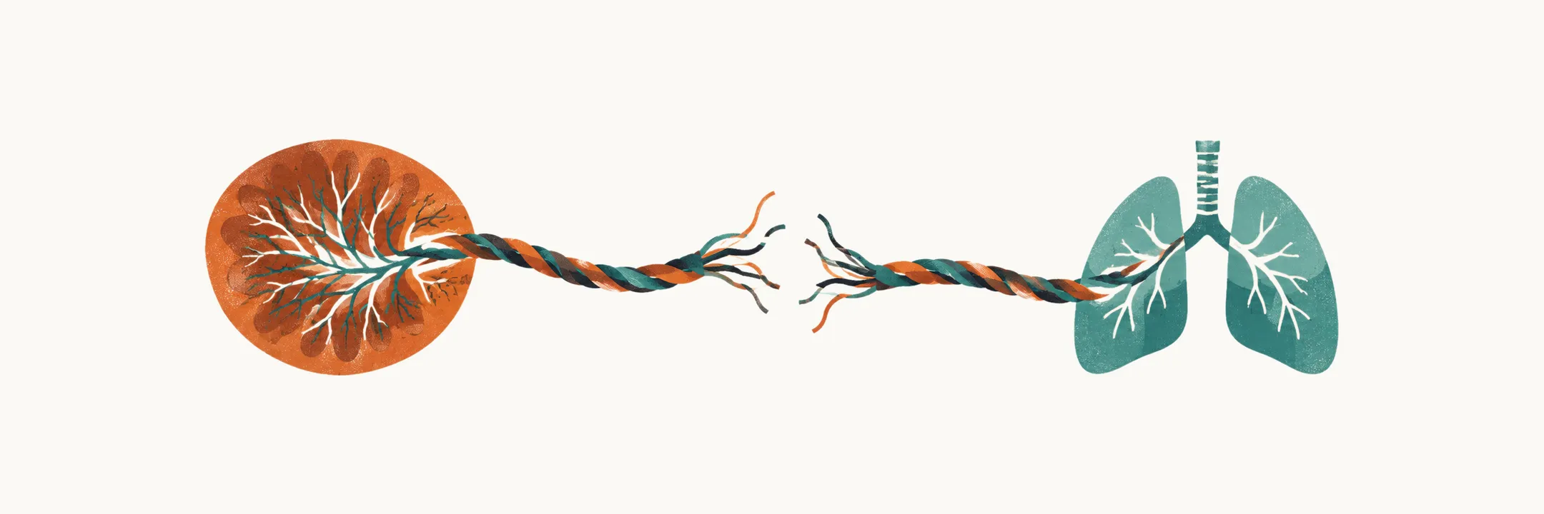
A preterm infant is a baby born before 37 completed weeks of gestation. Prematurity matters because birth removes placental support before the lungs, brain, gut, skin, immune system, respiratory control centers and cardiovascular transition are mature enough for independent life. The clinical problem is therefore not one disease but a mismatch between developmental and the demands of extrauterine life.
That mismatch is also why the complications arrive together rather than one at a time: they share the same substrate of immaturity, they interact with one another, and the same infant often carries several of them at once. They also follow a rough timeline after birth. The first hour tests breathing, temperature and circulation; the first weeks test the fetal circulation’s final changes, the fragile brain and the immature gut; infection and eye disease build up over the weeks of the admission; and some consequences appear only after the child has gone home. The obstetric side of prematurity — the causes of early delivery, preterm labor, preterm premature rupture of membranes (the membranes breaking before labor has begun), tocolysis (drugs used to delay labor) and antenatal corticosteroids — is a separate subject and is not covered here.
Where to start and where to go next
Start with Corrected Age and Clinical Risk in the Preterm Infant. It sets out how common preterm birth is, the prematurity bands from to , the mortality and morbidity gradient that falls as gestational age rises, why late-preterm infants are the easiest group to underestimate, and how corrected age — age counted from the expected due date rather than from birth — changes the interpretation of growth and development. That vocabulary is used throughout the other notes.
The first hour and the first days are dominated by stabilization and by the lung. The Golden Hour and NICU Care of the Preterm Infant follows the first hour: where the birth should happen and how the infant is transported, gentle respiratory support, protection from cold, cord clamping, monitoring and vascular access, and the environment and family experience of the neonatal intensive care unit (NICU). Respiratory Disease and Support in the Preterm Infant builds respiratory distress syndrome from a lack of surfactant (the substance that keeps small airspaces from collapsing) and an immature lung, then follows non-invasive pressure support, surfactant delivery, caffeine for pauses in breathing, air leak, and the transition to bronchopulmonary dysplasia, the chronic lung disease of prematurity.
Three notes follow the transitions of the first weeks. Patent Ductus Arteriosus in the Preterm Infant explains why the ductus arteriosus, the fetal vessel that lets blood bypass the lungs, often stays open; how the direction of flow through it either lowers oxygen saturation in the lower body or floods the lungs while drawing blood away from the organs; and how closure decisions trade pulmonary benefit against kidney and bowel toxicity. Intraventricular Hemorrhage and Brain Injury in the Preterm Infant covers bleeding from the germinal matrix, a fragile vascular region beside the ventricles, how the hemorrhage is graded and found by routine cranial ultrasound, the hydrocephalus that can follow, and the white-matter injury that predicts cerebral palsy. Nutrition and Necrotizing Enterocolitis in the Preterm Infant connects immature feeding, tube feeding, intravenous (parenteral) nutrition and central lines to necrotizing enterocolitis, inflammatory necrosis of the immature bowel, with its hallmark of gas in the bowel wall and its risk of perforation.
Two risks build over the admission. Infection and Sepsis in the Preterm Infant explains why prematurity is itself a risk factor, how early-onset and late-onset infection differ in source and organisms, and how the diagnostic and antibiotic decisions are made when the presentation is non-specific. Retinopathy of Prematurity in the Preterm Infant shows why retinal vessels that had not finished growing respond to oxygen and ischemia, how the disease is classified and screened for, when treatment is needed, and what oxygen targeting trades.
Long-Term Morbidity and Follow-Up in the Preterm Infant closes the sequence by following the child out of the unit: bronchopulmonary dysplasia, brain and eye outcome, growth and feeding, and how follow-up programmes monitor development using corrected age.